 Project leader
Holographic microscopy, also referred to as lensfree imaging, has been successfully explored by our team since 2005. Our most advanced device is a parallelized holographic video microscopy system which can monitor cell cultures in a 96-well microtiter plate. This optical system is based on an array of 12 x 8 image sensors, thus providing 96 densely integrated mini-microscopes. A mini-microscope is placed under every well of the transparent-bottomed microtiter plate and records the evolution of the cell culture over days or weeks. The array of video microscopes as well as dedicated software for viewing and processing image series are used in phenotypic screens to investigate and quantify viability, proliferation, morphology and motility of cells in 2D and in 3D.

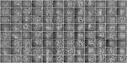
Overall view of a phenotypic screen onto clonal organoids (prostate acini) and clonal spheroids in a 96-well microtiter plate.
 Reference
Gabriel M, Balle D, Bigault S, Pornin C, Getin S, Perraut F, Block MR, Chatelain F, Picollet-D'hahan N, Gidrol X and Haguet V
Time-lapse contact microscopy of cell cultures based on non-coherent illumination.
Scientific Reports, 2015,
5: article 14532